The skin is the most extensive, complex living organ of the body, representing 12% of adult weight and 20% of the body’s water. It is a covering continuous with the mucous membranes at natural openings: it forms a fundamental anatomical and physiological barrier, the skin barrier, between the external environment and the internal environment. Age, race, sex, and the individual influence its structure and functions.
Author: JL Mathet – 2016
A complex and vital organ
It reflects the physiological state of the entire organism and helps maintain its biochemical and thermal homeostasis (i.e., equilibrium): thus, internal disturbances have cutaneous repercussions (hormonal diseases, infections, allergic states).
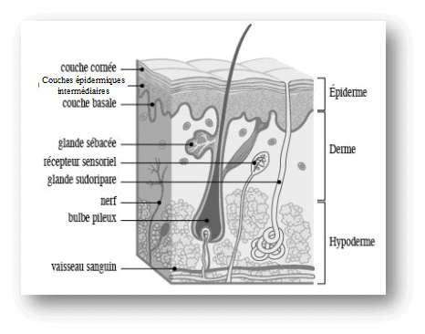
From the surface to depth, it consists of the epidermis, dermis, epidermal appendages (hair follicles, sebaceous and sweat glands, and lastly, the nail-like structures such as claws), hypodermis, and the cutaneous muscle (figure 1).
Its thickness varies from 0.5 to 5mm in dogs and varies according to the region of the body concerned, the breed, or the state of health (underlying diseases, deficiencies). It is thicker on the back and thinner in the ventral region and the distal parts of the limbs.
The pH
The pH of healthy dog skin is highly variable depending on multiple factors (breed, sex, area of the coat, environment, season). It is considered to be rather neutral to basic (from 7.4 to 8.5) unlike humans where the skin is more acidic. The use of topical treatments (shampoos, sprays, creams) should take this aspect into account to avoid any harmful variation in pH that may lead to skin barrier disruption and microbial proliferation.
The skin barrier
The different layers
The epidermis
It is an epithelium, that is, a non-mucous, keratinised, stratified surface covering, composed of layers of keratinocytes or corneous cells, without blood vessels. There are also non-epithelial cells with various functions: pigmentation with melanocytes, immune role with Langerhans cells and sensory role with Merkel cells and Meissner’s corpuscles.
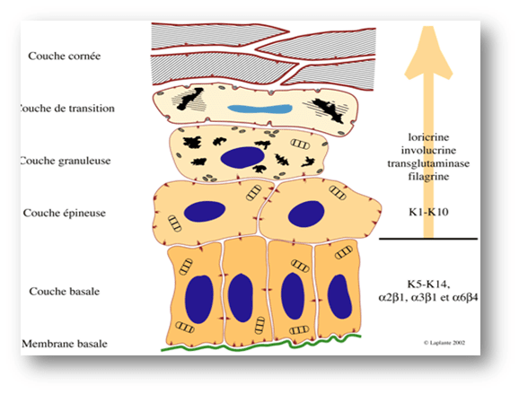
The epidermis is formed of 3 to 5 cellular layers in dogs: these layers are determined by the position, shape, morphology, and stage of differentiation of the keratinocytes.
From the depth to the surface: the basal layer is the germinative stratum, the spiny layer is a maturation compartment where attachments between the corneous cells (desmosomes) and lamellar bodies (lipid structures) form, the granular layer is a differentiation compartment, and finally the stratum corneum, which exfoliates during desquamation (figure 2).
The thickness of the dog’s epidermis varies from 20μm to 100μm (less than a tenth of a mm), and that of the stratum corneum from 5 to 20μm or up to 1500μm on the pads. It is thicker on the nose. In humans, the epidermis is also thicker.
The dermis
It is a vascularised connective tissue, formed of collagen and elastic fibres and an amorphous gel called intercellular matrix formed of hyaluronic acid, mucopolysaccharides, glycoproteins, and water.
Blood vessels organised by depth level, lymphatic vessels, nerve fibres, and numerous blood, inflammatory, and immune cells are found there.
The dermis has supporting and cushioning roles due to its elasticity, as well as immune and sensory roles. It also limits the diffusion of microbes and parasites that may have managed to cross the epidermis.
The hypodermis
It is a connective tissue formed of adipose (fatty) lobules separated by vascularised septa. It serves a lipid storage role, a thermal regulation role, and a mechanical protection role. It is the deepest tissue of the skin.
The concept of the “skin barrier”
The skin barrier is primarily represented by the stratum corneum, whose thickness varies from 5 to 1500 μm (depending on the location) and which provides the majority of the skin’s protective function but not solely, as various adhesion structures present in the living (stratified) layers of the epidermis are real units of intercellular communication that contribute to the physical and physiological cohesion of the barrier.
From the interlocking of angularly shaped corneocytes results the “brick wall” model, where the bricks are corneocytes, the cement is the intercellular lipid cement, and the intercellular junctions provide resistance and stability. This wall is gradually eliminated by enzymatic action in its superficial part during the desquamation process.
The association of structural proteins of corneocytes, keratins, and extracellular lipids constitutes a scaffold in close relation.
The initial “bricks and mortar” model, while still a basic reference, proved too rigid, and electron microscopy revealed that this “mortar” was a “sandwich” alternating a crystalline lipid phase and a fluid lipid phase organised in bilayers.
The barrier is a paradox in itself: it results from the superposition of layers of dead cells (corneocytes) yet whose functions are multiple and highly specialised, providing it with physical, chemical, and immune protective roles.
Its main roles
- mechanical : by cushioning shocks and resisting strains, also provided by conjunction with the dermis, by collagen and elastin fibres of the dermis, and by the hypodermis
- regulation of water exchanges : low permeability (lipophilic substances) to near-impermeability (hydrophilic substances), to water and most environmental agents
- protection against UV, toxins, and abnormal temperature variations (thermoregulation) and humidity by limiting the loss of water and electrolytes
- immune : defence against infectious agents by secreting antimicrobial molecules, and by the presence of resident microbial flora and the lipid surface film (antioxidant and pH role)
- thermoregulation : maintenance and regulation of temperature by the coat and the vascularisation of the dermis
- role sensory (itching, pain, temperature, touch) and social role, even if less pronounced than in humans
Cornification: formation of horn cells
This process, also called keratinisation, is the culmination of biochemical and morphological mechanisms that allow the transformation of the basal layer keratinocyte into a non-nucleated corneous cell, the corneocyte. It is therefore a permanent, regular, and harmonious renewal that physiologically lasts 3 to 4 weeks.
The successive stages of cornification are the germination then proliferation of keratinocytes in the basal layer, their maturation in the spiny layer, their differentiation in the granular layer then in the stratum corneum, and finally their exfoliation during the desquamation process, invisible physiologically to the naked eye.
Thus, the basal keratinocyte will proliferate and undergo a continuous, oriented programmed differentiation from the basal membrane towards the stratum corneum, involving complex protein and lipid syntheses, resulting in a dead corneous cell.
Any abnormal desquamation associates modifications of the hydrolysis of junctions between corneous cells, quantitative and qualitative disturbances of the lipids composing the epidermal film, and perturbations in the synthesis and secretion of enzymatic complexes. Its result is manifested by the abnormal formation of scales (clusters of corneocytes) in pathologies leading to seborrhoeic disorders or thickening of the stratum corneum (excessive keratinisation).
The surface lipid film
The sebaceous glands, sweat glands, and keratinocytes produce a protective lipid layer that covers the skin in mammals. The composition of glandular sebum differs from that of the inter-corneocyte film in humans as in animals. The former contains triglycerides, ester waxes, squalene, fatty acids, and cholesterol, the latter ceramides, free fatty acids, and cholesterol.
The lipid compartment of the epidermis includes the lipids attached to the outer surface of the corneal envelope and the lipids stored in the aligned lamellae of the intercorneocyte spaces. Lipid precursors are synthesised in the superficial spiny and granular layers and accumulated in secretory organelles (lamellar bodies or Odland bodies). They are then released into the intercorneocyte space and aggregate into parallel lipid sheets aligned with the hydrophobic layer of the corneal cell envelope.
Fur and coat
Fur
They are epidermal invaginations originating from a bud where the hair is manufactured and grows from the dermal papilla. Subsequently, this bud associated with the papilla will form the hair or follicle bulb: it is from this bulb that the entire hair follicle (i.e., the hair as a whole) will develop.
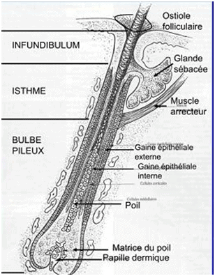
The hair follicle consists of three distinct parts from depth to surface: the producing part or bulb, the intermediate part or isthmus, and the outer part or infundibulum where the hair itself emerges. In dogs, several hair follicles can emerge from the same infundibulum, from 2 to 15; there are 100 to 600 hairs/cm². In cats, the hair density is more abundant, from 1000 to 2000 hairs/cm².
Additionally, two types of glands (sebaceous and sweat) open into it, as well as the arrector pili muscle. The latter allows the hair to stand up in certain behavioural sequences or to ensure better thermoregulation (insulating role of the coat).
The hair consists of a central part called the medulla filled with air for insulation, then the cortex which contains keratinised cells made of very hard keratin and pigments responsible for the hair’s colour, and finally the outer cuticle made of flattened, adherent keratin cells arranged like tiles. It is the cuticle that gives the coat its smooth feel. Longitudinally, from top to bottom, we distinguish the apex (the oldest part, the shaft with an outer zone (above the skin) and an inner zone (or infundibular)) and finally the root or bulbous part (figure 3).
The coat
Different types of hair are distinguished based on their size, diameter, and rigidity or conversely suppleness:
- the so-called primary (guard and cover) hairs are the largest, rigid hairs that give the coat its general appearance and colour; their distribution is generalised
- the secondary or undercoat, down/lanugo, or bristles, without sweat glands or arrector muscle, small, fine and flexible, they are also widespread
- the tactile (vibrissae, tylotrich hairs) with mechanical sensory functions, therefore associated with nerve fibres: they are found particularly on the face (eyelids, cheeks, lips) and on the forelimbs in cats (carpal organ)
Coats vary in length, colour and result from the proportion and distribution of each of these types of hair. The normal or mixed coat is close to the original wild coat (that of the wolf), medium-long as in the German Shepherd. The short coat can be coarse with mainly primary hairs (Beauceron, Rottweiler) or fine (short hair, mostly secondary hairs as in the Boxer, Doberman, Pinscher). The long coat can also be fine with mainly secondary hairs (Cocker, Spaniels, Yorkshire), or woolly (abundant down) as in the Poodle or Bichon. Finally, there are hairless dogs (Mexican Hairless Dog or Xoloitzcuintle). The length of a hair varies from 4 to 15 cm depending on the breed and species.
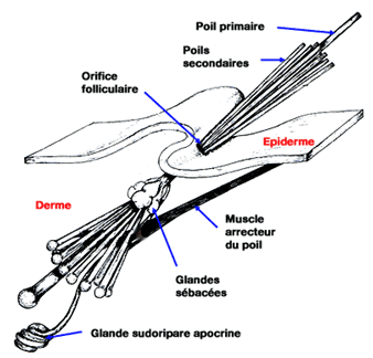
In cats, the coat is also composed of primary and secondary hairs. Likewise, there are short, long coats or hairless breeds (Rex and Sphinx). There are primary and secondary follicles grouped into units, in which the number of hairs varies. Secondary hairs are more common than in dogs, with a variable ratio of 1 primary hair for 10 to 20 secondary hairs, resulting in a particularly silky coat to the touch (figure 4). Vibrissae (‘whiskers’) and tylotrich hairs are particularly important in cats: their tactile sensory function is very advanced in the cat’s social life.
La colour and the length of coats are determined by the expression of genes passed down from generation to generation. This determinism is the basis of genetic tests in parents to predict the colour or length of the coat in a litter.
Hair cycle
Hair growth is cyclical, not continuous, and occurs in three successive stages called:
- anagen anagen or the so-called active growth phase
- anagen catagen or the transitional regression phase
- anagen telogen or resting phase, where the hair is dead but not immediately eliminated from the follicle
This growth is cyclical, and is not the same for every hair follicle: it influences the length of the hair and varies from one breed to another. It is influenced by external factors such as variations in day length (photoperiod), diet, external temperature (cold temperatures stimulate hair growth) and, of course, seasons (maximum growth). Internal factors specific to the individual also influence it, such as hormonal secretions or growth regulation factors. For example, thyroid hormones stimulate hair growth in anagen, adrenal hormones inhibit it. There are interracial and interindividual variations.
In dogs and cats, hair grows on average 0.3mm per day, particularly at the end of spring, with a slowdown in winter (more hairs in resting or telogen phase). Some breeds have prolonged growth (Poodles), requiring frequent grooming, others have a majority of dead hair (northern breeds).
Moults are periods of coat growth characterised by hair loss: growth is maximal and thus evacuates dead hair not yet fallen. Moults correspond to a progressive renewal of the coat in different areas, without synchronisation of one follicular group with another: this is called a ‘mosaic’ moult. Moults are permanent but vary in intensity according to the time of year. Thus, in Europe, the spring moult establishes a short, less dense summer coat, unlike the autumn moult which installs a long, dense winter coat.
There are successive coat aspects during the life of a carnivore: puppy coat is downy or woolly due to vertical hair implantation, which then gradually slopes. The coat of an aged dog becomes diffuse due to accelerated hair loss and decreased growth. It also becomes less shiny.
The coat primarily provides mechanical protection against external injuries, has an insulating and thermoregulatory function, and finally plays a role in the animal’s social and relational life (camouflage).
Skin glands
In dogs, sebaceous glands producing sebum and sweat glands producing sweat are distinguished.
Sebaceous glands
In dogs, sebaceous glands open into the hair canal and are closely associated with the hair follicle, unlike in humans where the duct opens directly onto the skin’s surface. Sebum is a lipid-rich secretion (cholesterol, glycerides and waxes) that contributes to the surface lipid film with epidermal lipids, thus helping to maintain an effective skin barrier. It provides microbial protection, skin waterproofing, and supplies skin suppleness and coat gloss.
Specific sebaceous glands are found on the eyelids, in the ear canal, on the tail (supra-caudal gland), chin, anal sacs (‘anal glands’) and around the anus (circumanal glands).
Sebum production is regulated by numerous endocrine, nutritional, and genetic factors. When the quantity or quality of sebum is disrupted, dry or oily seborrhea may occur, leading to unpleasant and strong odour, greasy or dull coat, dandruff (‘flakes’) production and microbial proliferation. This results in kerato-seborrheic conditions, common skin issues in dogs with chronic infectious complications (photos 1 and 2).
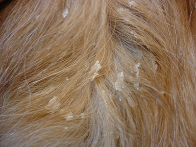
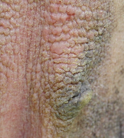
Sweat glands
Sweat glands allow partial regulation of the dog’s internal temperature by perspiration, linked to sweat evaporation. This phenomenon is less pronounced than in humans; it is mainly the dog’s breathing that allows thermoregulation. The odorous molecules contained in sweat allow social recognition.
Conclusion
The skin and coat are thus fundamental elements of protection, interaction, and communication for the animal itself but also between individuals. The variations in appearance and renewal of the fur are significant in dogs and cats due to racial genetic selections and standards of beauty. The skin also reflects the health of the pet, and its dermatological and cosmetological care is essential for every owner. Canine and feline dermatology is indeed a superb and fascinating specialty, which maintains close links with many other medical disciplines.
To learn more…
- Practical Guides to Canine and Feline Dermatology by Drs E. GAGUERE and P. PRELAUD, Ed KALIANKIS – MERIAL, 2006.
- Practical Guide to Dermocosmetics by Dr E. BENSIGNOR, Ed MED’COM, 2016.
- Knowing the skin of the dog and its diseases. Drs E. BENSIGNOR and C. HADJAJE, Ed MED’COM, 2013.
Recherches Connexes
How many hairs does a cat have, supracaudal gland cat, sebaceous gland diagram, animal skin, flaky dog skin, cats, undercoat, device, grooming, well-being